
1. What is a Demodex Mite Test?
Demodex mites are tiny mites that live in human hair follicles and sebaceous glands. They can be found even on the skin of healthy adults, and their presence alone does not indicate a disease.1
However, when Demodex mites become excessively numerous, they can cause inflammatory lesions such as mid-facial redness, rough scaling, papules, and pustules. Their association with rosacea is particularly well-known.2
The Demodex mite test evaluates for an overgrowth of Demodex mites in such lesions. At our clinic, we utilize polarized light and UV dermoscopy.
2. Findings in Polarized Dermoscopy
Check whether Demodex tails and Demodex follicular openings are repeatedly observed in multiple pores within the lesion area.3,4
- Demodex mite tail: Refers to a white or yellowish-white, gelatinous, thread-like structure protruding outside the pore.
- Demodex pores: Refers to dilated pores filled with gray or light brown plugs, with the surrounding area appearing red.
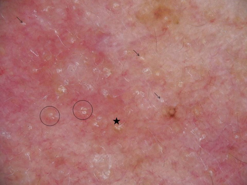
Arrows indicate Demodex tails, asterisks indicate Demodex openings, and circles indicate thread-like structures emerging from the follicles. Source: Friedman et al. Dermatol Pract Concept. 2017;7(1):35-38, Figure 3. Original · CC BY 4.0
3. Findings in UV Dermoscopy
Check the density of bright blue fluorescent dots. The density of these dots tends to be proportional to the number of Demodex mites.5
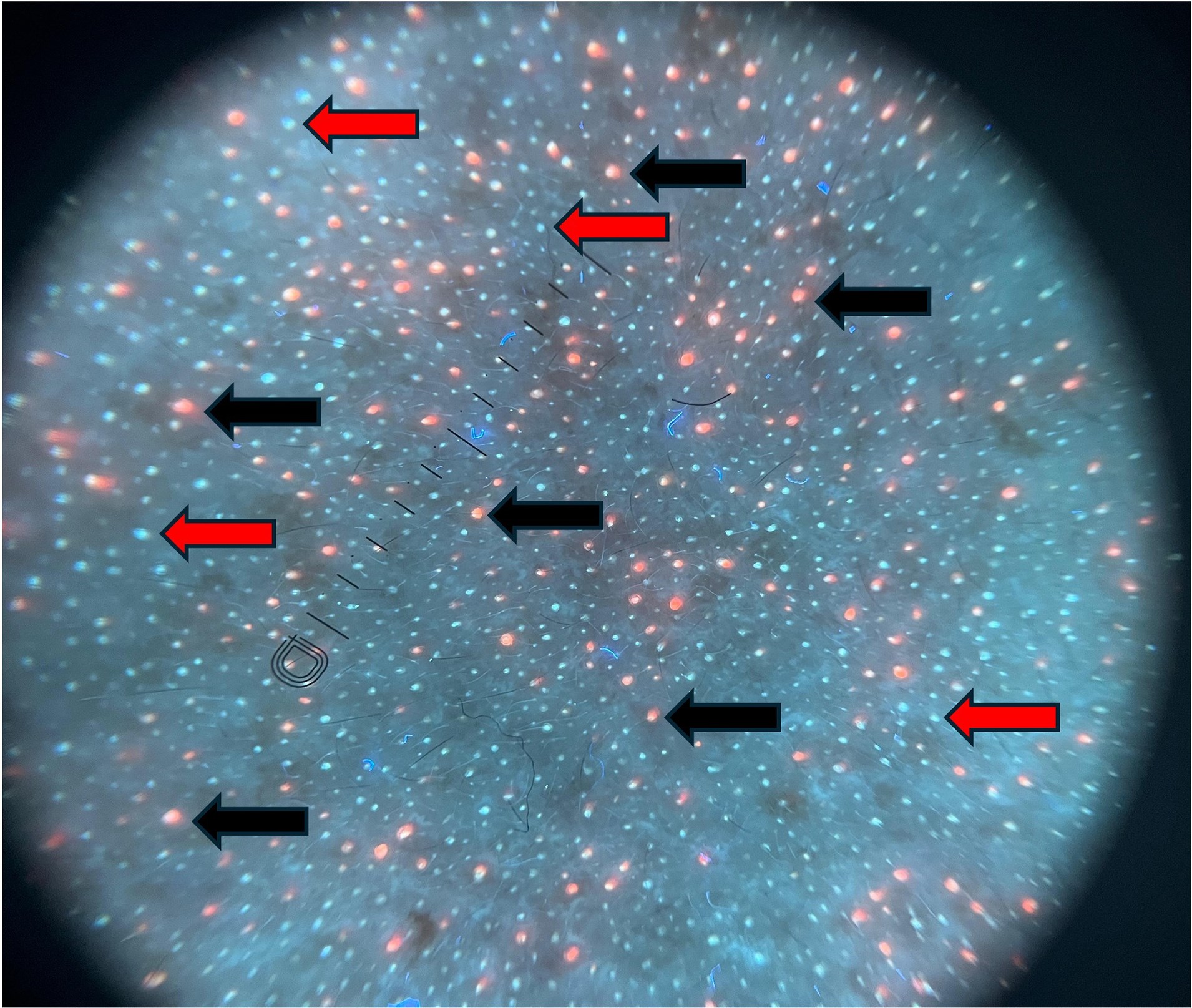
Black arrows indicate bright blue fluorescent dots, and red arrows indicate orange fluorescent dots. Source: Ünal et al. Clin Exp Dermatol. 2026, Figure 1. Original · CC BY 4.0
Frequently Asked Questions
Q. Does everyone have Demodex mites?
A. They can be found on the skin of most adults. The issue is not the presence of Demodex itself, but when they are observed in excessive numbers alongside inflammatory lesions.
Q. Is the Demodex mite test painful?
A. It is not painful. We observe the skin under magnification using polarized and UV dermoscopy, and no samples are collected.
Q. Can I get the test results immediately?
A. Results can be confirmed immediately in the consultation room. However, test findings are interpreted in conjunction with symptoms, lesion distribution, and treatment response.
Q. Is the test repeated after treatment?
A. If necessary, we re-examine the same area to check for changes in Demodex-related findings.
Q. How much does the examination cost?
A. This is routinely checked during consultation when necessary, and there is no separate testing fee.
References
- Chen W, Plewig G. Human demodicosis: revisit and a proposed classification. Br J Dermatol. 2014;170(6):1219-1225.
- Chang YS, Huang YC. Role of Demodex mite infestation in rosacea: A systematic review and meta-analysis. J Am Acad Dermatol. 2017;77(3):441-447.e6.
- Friedman P, Sabban EC, Cabo H, et al. Usefulness of dermoscopy in the diagnosis and monitoring treatment of demodicidosis. Dermatol Pract Concept. 2017;7(1):35-38.
- Parra-Cares J, Meza-Romero R, Ibáñez S, et al. Dermoscopy as a clinical tool for the diagnosis of demodicosis: a retrospective intrapatient case-control study. Arch Dermatol Res. 2024;316(8):509.
- Ünal E, Yücel MB, Yiğit M, et al. Detection of Demodex in Rosacea: Standardized Skin Surface Biopsy versus Ultraviolet Dermoscopy. Clin Exp Dermatol. 2026. doi:10.1093/ced/llag100.