1. What is a dermoscopy examination?
Dermoscopy is a non-invasive examination that magnifies the skin to observe its structure and fluorescence.
The dermoscope we use supports both polarized and UV modes. Viewing the same skin with both methods allows us to assess structure and fluorescence separately.
2. What’s the difference between polarized light and UV?
2.1 Polarized Dermoscopy
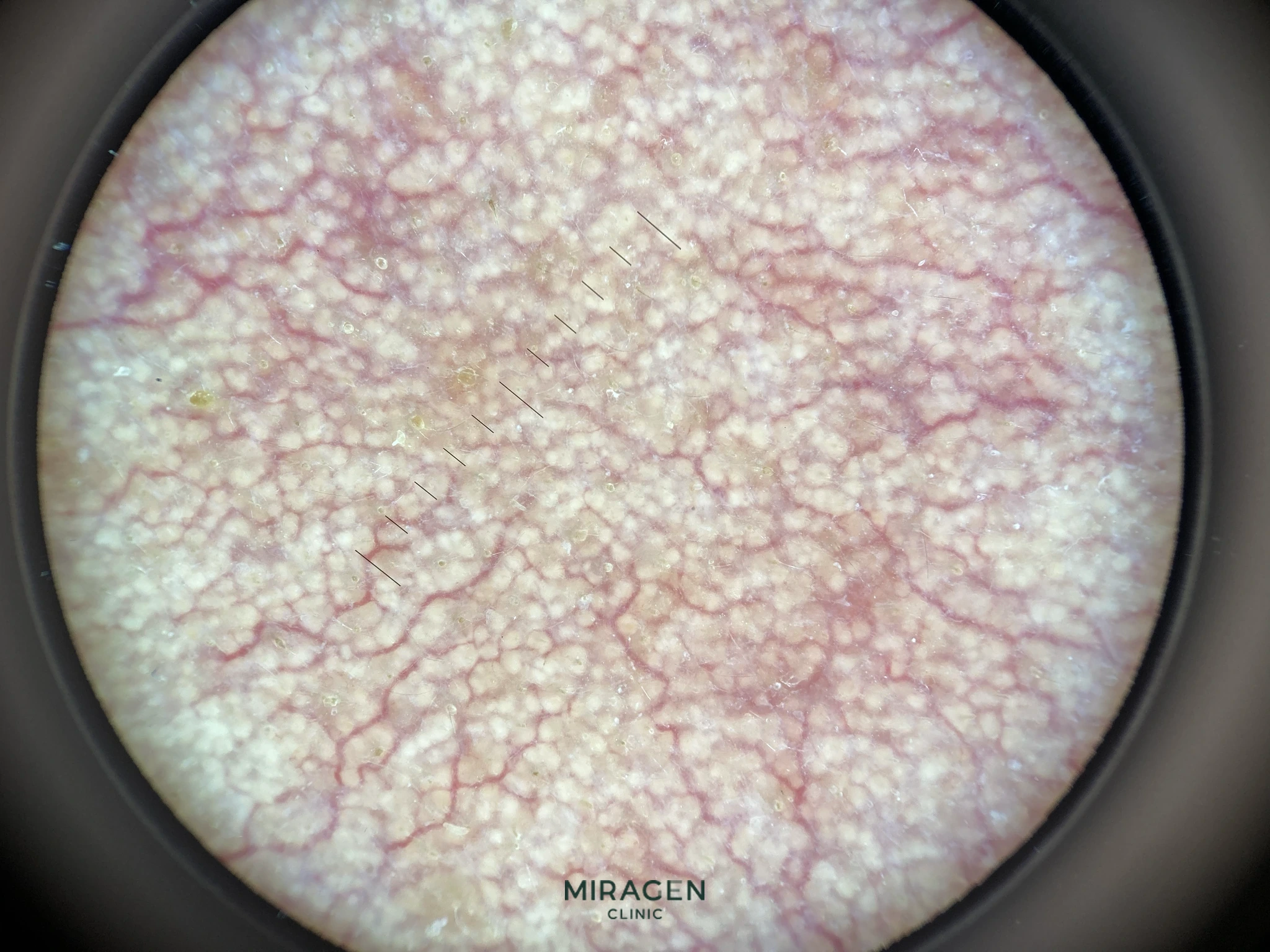
Polarized light reduces reflections from the skin surface, making it easier to observe the skin’s surface, pigments, blood vessels, and pores. At our clinic, we use it to evaluate excessive Demodex proliferation in areas of dermatitis or folliculitis, and to determine whether redness is caused by dilated blood vessels or dermatitis.1
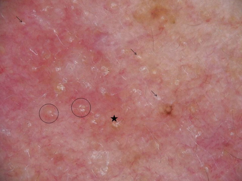
Arrows indicate Demodex tails, asterisks indicate Demodex openings, and circles indicate thread-like structures emerging from the follicles. Source: Friedman et al. Dermatol Pract Concept. 2017;7(1):35-38, Figure 3. Original · CC BY 4.0
2.2 UV Dermoscopy
UV enables us to observe the color, location, and distribution of fluorescence emitted from pores and skin.2 At our clinic, it is utilized to evaluate the excessive proliferation of acne bacteria, Demodex, and Malassezia in lesion areas.
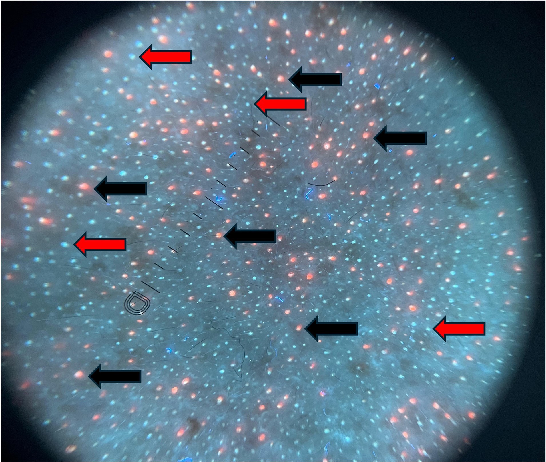
Black arrows indicate bright blue fluorescent dots, and red arrows indicate orange fluorescent dots. Source: Ünal et al. Clin Exp Dermatol. 2026, Figure 1. Original · CC BY 4.0
3. Test Method
The exam is performed immediately in the consultation room. We place the device on or close to the skin and switch between polarized and UV modes as needed. It’s painless, and you can check the results right away.
4. Our Clinical Application
At our clinic, we utilize dermoscopy examinations to identify the cause in areas affected by redness, dermatitis, and folliculitis.
- Redness: With polarized light, we check whether telangiectasia is clearly visible.3 If dilated vessels are not clearly seen, we consider treating dermatitis first rather than Excel V.
- Dermatitis · Folliculitis: We observe Demodex tails and pore structures under polarized light, and check the distribution of red and blue dots using UV light. If signs of excessive microbial growth are observed, we select the appropriate topical treatment based on the type. Detailed information is provided on the Demodex Examination page.
Frequently Asked Questions
Is the test painful?
It is not painful. Because it is an examination that magnifies and observes the skin, injections or blood draws are not required.
Can I know the results right away?
You can check them immediately in the consultation room. However, the results are interpreted together with your symptoms, medical history, and skin condition.
How is it different from a Wood’s lamp exam?
A Wood’s lamp exam lets you see the fluorescence distribution over a wide area at a glance. UV dermoscopy magnifies each individual pore to assess the location and distribution of fluorescence.
Is there a separate cost for the test?
No. It’s a basic check we perform as needed during your visit, so there is no additional charge.
References
- Lallas A, Argenziano G, Apalla Z, et al. Dermoscopic patterns of common facial inflammatory skin diseases. J Eur Acad Dermatol Venereol. 2014;28(5):609-614.
- Pietkiewicz P, Navarrete-Dechent C, Togawa Y, et al. Applications of Ultraviolet and Sub-ultraviolet Dermatoscopy in Neoplastic and Non-neoplastic Dermatoses: A Systematic Review. Dermatol Ther (Heidelb). 2024;14(2):361-390.
- Micali G, Dall’Oglio F, Verzì AE, et al. Treatment of erythemato-telangiectatic rosacea with brimonidine alone or combined with vascular laser based on preliminary instrumental evaluation of the vascular component. Lasers Med Sci. 2018;33(6):1397-1400.